

Konus Prepared Slides: Pathological Tissues 1


- ✅ Set of 10 Konus prepared slides with human pathological tissue specimens, ideal for learning and teaching in biology and medicine.
- ✅ Includes slides representative of diseases such as tuberculosis, liver cirrhosis, splenic leukemia, pneumonia, malaria, and colon cancer.
- ✅ Allows observation of cellular and structural alterations caused by specific diseases on real tissues, stimulating diagnostic understanding.
- ✅ Useful visual support for health science students and faculty in histology and pathology exercises.
- ✅ Professional-quality product, also ideal for laboratories and enthusiasts of microscopy and human pathology.


Full description
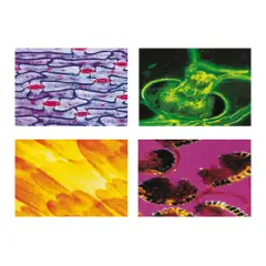
Konus brand "Pathological Tissues 1" prepared slides offer a unique opportunity to explore the world of pathological changes in human organs and tissues. Each slide contains a sample of human tissue that has undergone changes due to a specific pathological condition. This set is especially useful for students of biology, medicine, and health sciences who wish to deepen their understanding of diseases and how they affect the structure and function of organs in the human body.
Contents of prepared slides:
Pulmonary tuberculosis
A lung tissue specimen showing signs of tuberculosis. Tuberculous granulomas and other cellular changes caused by Mycobacterium tuberculosis infection can be seen.Cirrhosis of the liver
A section of liver affected by cirrhosis. Liver cell damage, fibrosis formation, and loss of normal liver structure due to chronic damage are observed.Leukemia of the spleen
A spleen specimen showing the changes caused by leukemia. Blood cancer cells can infiltrate splenic tissue, altering its structure.Pneumonia
A section of lung affected by pneumonia, showing inflammation and infiltration of inflammatory cells (such as neutrophils and macrophages) in the lung tissues, typical of a bacterial or viral infection.Malaria (blood)
A blood smear showing the presence of Plasmodium responsible for malaria. Alterations in red blood cells caused by the Plasmodium parasite are visible, along with its developmental stage.Colon cancer
A sample of colon tissue affected by a tumor. Tumor cells may appear disorganized from the normal arrangement of epithelial tissue, and neoplasm formations are visible.Rheumatoid arthritis
A joint specimen showing signs of rheumatoid arthritis. Inflammatory cells are evident, with joint tissue damage such as synovitis and loss of articular cartilage.Cystic fibrosis (lung)
A lung specimen showing the changes caused by cystic fibrosis. Obstruction of the respiratory ducts by thick mucus and lung tissue damage caused by the genetic disease can be seen.Atherosclerosis (artery)
An artery specimen showing the atherosclerotic plaques formed on the arterial walls. Structural changes and deposition of lipids and fibrosis in the vessel walls are evident.Myocardial infarction
A section of heart showing the damage caused by a myocardial infarction (heart attack). Necrotic areas of heart tissue and the body's inflammatory response to cell death are observed.
With these slides, it is possible to better understand the pathogenetic mechanisms underlying many pathological conditions, as well as to develop a deeper understanding of the microscopic diagnosis of diseases.
This article can be found at pg. 193 Of our primary and secondary catalog